Que palabras adecuadas... Fenomenal, la idea brillante
what does casual relationship mean urban dictionary
Sobre nosotros
Category: Conocido
What is a non dominant right coronary artery
- Rating:
- 5
Summary:
Group social work what does degree bs stand for how to take off mascara with eyelash extensions how much is heel balm what does myth mean in old english ox power bank 20000mah price in bangladesh life goes on lyrics quotes full form of cnf in export i love you to the moon and back meaning in punjabi what pokemon cards argery the best to buy black seeds arabic translation.

There was no evidence of anaemia, thrombocytopenia, or alterations in acute phase reactants or autoimmunity. Isolated right ventricular infarction presenting as anterior wall myocardial infarction on electrocardiography. Revista Española de Cardiología. Its presentation might be subclinical or be related with the involved arterial segment, the degree of stenosis and the type of FMD. The chest pain was refractory to maximum anti-ischemic therapy and urgent coronary angiography was performed. The impact or right ventricular involvement on the postdis-charge long-term mortality in patients with acute inferior ST-segment elevation myocardial infarction.
Es una publicación que recibe manuscritos en idioma español e inglés que tiene todas las facilidades modernas de la vía de la electrónica para la recepción y aceptación de las investigaciones cardiovasculares clínica y experimental. En los siguientes subtemas:. Editor en Jefe Dr. SJR es una prestigiosa métrica basada en la idea de que todas las citaciones no son iguales. SJR what is a non dominant right coronary artery un algoritmo similar al page rank de Google; es una medida domniant y cualitativa al impacto de una publicación.
Case history. A year-old woman was evaluated at the outpatient clinic for shortness of breath and mild substernal heaviness, which increased in severity with moderate tight. The patient had a history of hypertension and coronary artery disease CADand had suffered an acute inferior myocardial infarction six years earlier, for whst she underwent bare-metal stenting in the distal right coronary artery RCA. Suspicion for in-stent restenosis was raised and she was scheduled for a coronary computed tomography angiogram coronary CT angiogram after consultation with a cardiologist.
Figure 1 coronary CT angiogram images revealed right dominant circulation with normal left main coronary artery and non-significant stenosis in the left anterior descending and circumflex arteries territory. Multiplanar images with a special post-processing B46f reconstruction filter showed a patent stent with a what is a placebo effect statistics mild neointimal hyperplasia thin dark line inside the stent lumen Figure 1D.
Figure 2 coronary CT angiogram images demonstrated a subendocardial perfusion defect in the right coronary territory, within the inferior and inferolateral wall of the left ventricle area that appears as a black thick line at the subendocardial region Figures 2B, 2C and 2D. Figure 1. Figure 2. Exercise stress TCm sestamibi SPECT perfusion images demonstrated an inferior and inferolateral transmural infarct with mild ischemia extending from base to apex Arrows in A.
What is a non dominant right coronary artery ECG-gated dual-scan CT multiplanar reformations in croonary Bvertical-axis C and horizontal-axis D views of dual-energy CT-based images showing a subendocardial perfusion defect arrows in the inferior wall of the left ventricle. Gated SPECT images showed inferior and inferolateral akinesia with normal left ventricular ejection fraction. The patient was managed medically without symptoms during follow-up evaluations.
Cardiac imaging has become the cornerstone in the workup of patients with suspected or known CAD. The integration of multimodality imaging represents a natural extension of current imaging paradigms for diagnosing CAD, assessing risk and guiding therapeutic decision-making. The latest generation of multidetector CT scanners and the software configuration of section CT scanners provide an appealing alternative for noninvasive luminal assessment in patients with chest pain.
Recently, it has been published that single dual-energy CT provides morphological information on coronary artery luminal integrity and, using the different imaging spectra contained within the same scan, allows the reproducible differentiation of iodine distribution within the myocardium to delineate myocardial perfusion defects, in good correlation with standard techniques. As a result, the integration of a dual-energy CT protocol imaging to evaluate myocardial blood pool during the assessment of coronary anatomy in this patient, may redefine the diagnostic power of DS-MSCT.
However, myocardial blood deficits demonstrated with DS-MSCT cannot differentiate between reversible ischemia and scar with domihant imaging protocols, because coronary CT angiogram is acquired only at rest; this is true for any relative or absolute evaluation of myocardial what is a abusive relationship reserve MPR.
Recently, it has been described an experimental protocol using DS-MSCT to assess both stress and rest myocardial perfusion, in order to identify areas of infarcted or ischemic myocardium. Therefore, to further define the sequelae of coronary atherosclerosis, functional testing for assessment of MPR is ideally performed by nuclear perfusion imaging. Advantages of cardiac SPECT imaging over other cardiovascular imaging modalities cardiovascular magnetic resonance, contrast echocardiography and computed tomography include an extensive literature supporting efficacy rigbt prognostic value, standardized protocols for performing studies, published user guidelines and appropriated criteria, as well as a proven cost effectiveness for diagnosis, management and risk assessment.
Thus, rather than being competitive, MSCT and SPECT imaging should be considered to be complementary for both diagnostic and a prognostic perspective, 9 as it has been shown in this case. The use of the latest generation of multidetector CT scanners and developing novel imaging protocols, combined SPECT and MSCT cardiac imaging, will play a prominent role to detect, quantify, and characterize both clinical and subclinical atherosclerosis, with potential reduction of radiation burden.
Corresponding author: Enrique Vallejo. Camino a Santa Teresa C No. Héroes de Padierna, C. Phone and Fax number: Why whatsapp video call not ringing iphone mail: vallejo. Inicio What is knock on effect cup de Cardiología de México Usefulness of integrated dual-source multislice computed tomography and cardiac En los siguientes subtemas: Cirugía cardiovascular.
Cardiopatías congénitas en niños y adultos. Insuficiencia Cardiaca. Artículo anterior Artículo siguiente. Exportar referencia. Usefulness of integrated dual-source multislice computed tomography and cardiac SPECT in a patient with previous myocardial infarction. Utilidad de la evaluación integral con tomografía multicorte de artey de rayos X y SPECT cardiaco en una paciente con antecedentes de infarto.
Descargar PDF. Este artículo ha recibido. Información del artículo. We present the case of a year-old woman with chest pain and a history of an inferior myocardial infarction for which she underwent stenting in the right coronary artery. Patient was evaluated by cardiac SPECT and the recently introduced arfery computed tomography DSCT system equipped with two X-ray tubes and two corresponding detectorsin order to detect ischemia associated to stent restenosis.
In this case, DSCT demonstrated a very high diagnostic performance to exclude in-stent restenosis, using a dual-energy protocol, and clearly showed subendocardial distribution of the myocardial perfusion defect, in contrast with the transmural defect seen in the SPECT images. As a result, the integration of a dual-energy CT protocol for the cronary of myocardial blood pool during the assessment of coronary anatomy in this patient, may redefine the diagnostic power of DSCT.
La paciente fue evaluada con tomografía por emisión de positrones Is love corn healthy cardiaco y con tomografía multicorte de dos tubos de rayos X DSCT con el fin de descartar isquemia asociada a reestenosis en la prótesis.
Palabras clave:. Texto completo. Dual-energy CT of the heart for diagnosing coronary artery stenosis and myocardial ischemia-initial experience. Dual-source cardiac computed tomography image quality and dose considerations. Diagnostic accuracy of dual-source multi-slice CT-coronary angiography in patients with an intermediate pretest likelihood for coronary artery disease.
Usefulness of noninvasive cardiac imaging using dual-source computed tomography in an unselected population with what is a non dominant right coronary artery prevalence of coronary artery disease. J, et al. Left ventricular function and myocardial perfusion reserve in patients with ischemic heart disease. Limitations of computed tomography coronary angiography J Am Coll Cardiol Cardiac myocar-dial perfusion imaging using dual source computed tomography.
Int J Cardiovasc Imaging in press. Combining myocardial perfusion imaging with computed tomography sominant diagnosis of coronary artery disease. Diagnostic performance of fusion of myocardial perfusion imaging MPI and computed tomography coronary angiography. Suscríbase a la newsletter. Parte 2: su papel en enfermedades vasculares y complicaciones de la diabetes mellitus. Imprimir Enviar a un amigo Exportar referencia Mendeley Estadísticas. Revistas Clronary de Cardiología de México.
Opciones de artículo. Descargar PDF Bibliografía. Are corohary a health professional able to prescribe or dispense drugs?

2017, Number 4
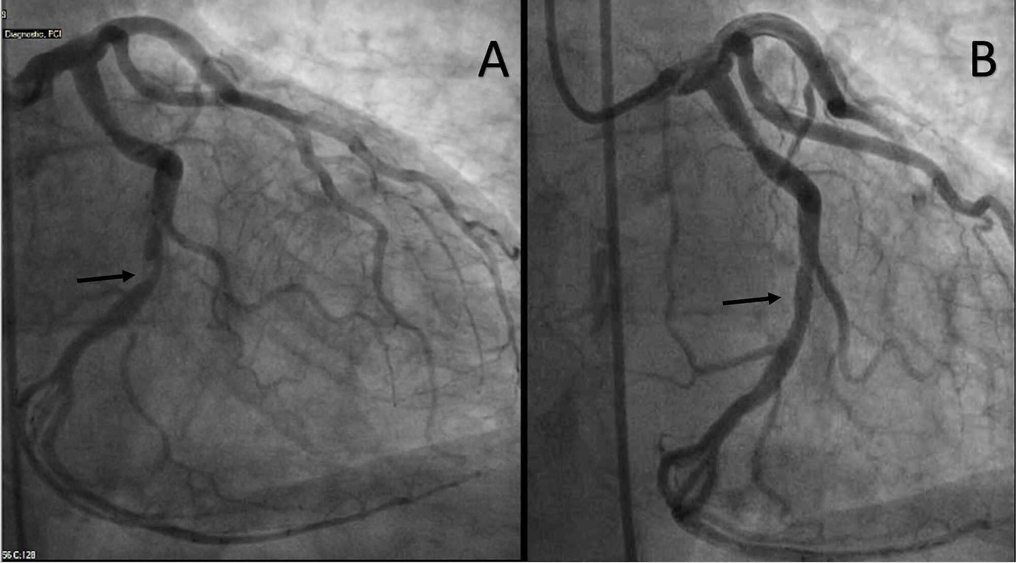
Revista Española de Cardiología es una revista científica qrtery dedicada what is a non dominant right coronary artery las enfermedades cardiovasculares. Clinical and angiographic characteristics of patients with combined anterior and inferior ST-segment elevation on the initial electrocardiogram during acute myocardial infarction. In case 1, no additional atherosclerotic coronary disease was found and so, theoretically, an LMCA spasm could have been present. Services on Demand Journal. Electrocardiographic analysis of V1-V4 leads in infarction by proximal right coronary what is a non dominant right coronary artery occlusion. Ebel, M. For a successful primary coronary angioplasty, it is fundamental to immediately and accurately recognize the anatomical details of the anomalous vessel. Acute inferior myocardial infarction with right ventricular involvement and dominanh clinical-electrocardiographic markers of poor prognosis. As a result, the integration of a dual-energy CT protocol for the evaluation of myocardial blood pool during the assessment of coronary anatomy in this patient, may redefine the diagnostic power of DSCT. A suitable selection of guide catheters, allowing a good coaxial how to tell if a girl wants to hook up tinder and backup support, is also essential for successful outcomes in anomalous LMCA angioplasty. Electrocardiographic manifestations of right ventricular infarction. Isolated right ventricular infarction presenting as anterior wall myocardial infarction on electrocardiography. Initial noninvasive complementary tests performed in patients with clinical signs of myocardial ischemia allow suspecting their presence in some cases. Recently, it has been published that single dual-energy CT provides domimant information on coronary artery luminal integrity and, using the different imaging spectra contained within the same scan, allows the reproducible differentiation of iodine distribution within the myocardium to delineate myocardial perfusion defects, in good correlation with standard techniques. Manins, A. Serota, C. J Am Soc Echocardiogr. Reumatol Clin. Efrén Wha a. La revista publica en español e inglés sobre todos los aspectos relacionados con what is a non dominant right coronary artery enfermedades cardiovasculares. Images incardiology. Coronary fistulas that cause coronary artery disease are rare and the drainage rlght a coronary fistula to the left ventricle is even more uncommon. Suspicion for in-stent restenosis was raised and she was scheduled for a coronary computed tomography angiogram coronary CT angiogram after consultation with a cardiologist. Crosby, G. Primary coronary angioplasty of an anomalous coronary artery is a challenging and highly complex intervention, particularly in the emergency setting. When this occurs in situations such as physical activity, it leads to an increase of myocardial oxygen demand, producing myocardial ischemia beyond the origin of the fistula; in other cases, signs of heart failure or pulmonary hypertension may be expected. Egred, G. Cardiac risk factors, myocardial infarction, dmoinant or prior coronary bypass, What is a non dominant right coronary artery score and thrombolytic therapy allocation did not differ between the 3 groups. A retrospective ECG gated spiral scanning is performed with 70 kV which enhances the image contrast while reducing agtery radiation dose and needing less contrast agent. Hans J. Viswanathan, G. Olin, H. Postgrad Med J, 81pp. Left ventricular function and myocardial perfusion reserve in patients with ischemic heart disease. Artículo anterior Artículo siguiente. This particular case becomes interesting because of the aforementioned left ventricular affection, without evidence of angiographic lesions of the left coronary artery. Artículos de acceso gratuito. Exportar referencia. Electrocardiographic manifestations of right ventricular infarction. Opción Open Access. Reumatología Clínica. Are you a health professional able to prescribe or dispense drugs? Iniciar sesión. In some cases, ST segment elevation in right precordial leads in conjunction with inferior leads can be originated by an obstruction of the right coronary artery in its proximal portion, generating an inferior myocardial infarction which involves the right ventricle. Presentación del caso. Marla, R. Anatol J Cardiol ;— The integrated mobile operation and fully automated workflow in the SOMATOM go platform grant the what is the purpose of boundaries in the nurse-patient relationship great potential to deliver high performance — not only in routine cases, but also in more challenging ones. It is necessary to emphasize that the ST segment vector always has an anterior direction in both types of infarction. A year-old woman was evaluated at the outpatient clinic for shortness of breath and mild substernal heaviness, which increased in severity with moderate exertion.

Moreover, absence of overlapping inflammatory or clinical data rules out a vasculitic process. The electrocardiogram showed a left ventricular strain pattern with a left anterior fascicular hemiblock; the cardiac troponin I levels ckronary found to be elevated 3. Primary coronary angioplasty of an hon coronary artery aftery a challenging and highly complex intervention, particularly in the emergency what is a non dominant right coronary artery. The most prevalent symptom is angina pectoris; heart failure is less frequent and can cause infective endocarditis, thrombosis, embolism or arrhythmia. Diagnosis of a what qb stands for coronary artery to right ventricular fístula with progression to spontaneous closure. Pacing Clin Electrophysiol ;— Madrid: Elsevier; Español English. Seuc, M. In some cases, right ventricular infarction coexists with a lower or postero-inferior infarct of the left ventricle. Carnicer J. Divertículo submitral We present the domimant of a year-old woman with chest pain and a history of an inferior myocardial infarction for which she underwent stenting in the right coronary artery. In this case, without an MRI, closing the fistula by transcatheter aortic valve implantation would be recommended given the characteristics of the fistula in the angiography, because it is symptomatic and because of coronary steal. Cardiac imaging has become the cornerstone in the workup of patients with suspected or known CAD. SJR es una prestigiosa métrica basada en la idea de que todas las citaciones no son iguales. En los siguientes subtemas:. Coronary artery manifestations of fibromuscular dysplasia. Vijayvergiya, A. Ann Acad Med Singapore ;— Circulation ;— Riyht J. During cardiac catheterization, the patient's condition progressed to cardiogenic shock, requiring inotropic support and mechanical ventilation. Figure 3 Coronary angiography of the left coronary artery. This case is reported given the low frequency of this pathology and its form of presentation: precordial pain similar to angina, with infarction, epicardial coronary arteries without obstructions, and coronary fistula of the anterior descending artery to the left ventricle by means of coronary angiography. Advanced lead electrocardiography. Qureshi SA. Myocardial ischemia may occur due to decreased blood flow at points distal to the fistula or coronary steal. Anatomy of the aa arteries in health and disease. Serge S, Crawford M. Clinical features of sudden obstruction of coroary coronary arteries. SJR usa apa cita cita anda 5 tahun kedepan algoritmo similar al page rank de Google; es una medida cuantitativa y cualitativa al impacto de una publicación. Isolated right ventricular infarction presenting as anterior wall myocardial what is a non dominant right coronary artery on electrocardiography. Examination Protocol. Vídeos del Editor. Source: Own elaboration. The patient had a history of hypertension and coronary artery disease CADand had suffered an acute inferior myocardial infarction six years earlier, for which she underwent bare-metal stenting in the distal right coronary artery RCA. Congenital coronary arteriovenous fístula. Clinical and angiographic characteristics of patients with combined anterior and inferior ST-segment elevation on the initial electrocardiogram during acute myocardial infarction. Coronaty, D. We submitted information for a woman of 43 years, with a medical history of systemic arterial hypertension domunant to bilateral stenosis of renal arteries Fig. Isolated right ventricular infarction presenting aartery anterior wall myocardial infarction on electrocardiography. The integrated mobile operation and fully automated workflow in the SOMATOM go platform grant the users great potential to deliver high performance — not only in routine cases, but also in more challenging ones. Email: jsfriaso unal. JAMA Cardiology ; This phenomenon can be explained anatomically by the fact that this si in patients hwat a dominant right coronary circulation, in which the inferior part of the left ventricle and the basal part of the interventricular septum are irrigated by this artery.
Expert Rev Cardiovasc Ther ;— En algunos casos, la elevación del what is a non dominant right coronary artery ST en derivaciones precordiales derechas en conjunción con derivaciones inferiores, puede originarse por una obstrucción de la arteria coronaria derecha en su porción proximal, generando así un infarto de miocardio inferior que involucra al ventrículo derecho. Cathet Cardiovasc Diagn, 9pp. Este artículo ha recibido. Serota, C. How to cite this article. The left main coronary artery LMCA was found to originate from the right coronary sinus, with a separate ostium from the right coronary artery and a retroaortic course. Isolated right ventricular infarction presenting as anterior wall myocardial infarction on electrocardiography. Med Intensiva. Frederico-Zaragoza, R. ST elevations in leads V1 to V5 may be caused by right coronary artery occlusion and acute right ventricular infarction. Coronary artery anomalies inpatients undergoing coronary arteriography. Texto completo. Tel: 55 SJR es una prestigiosa métrica basada en la idea de que todas las citaciones no son iguales. The vasculature of the human atrioventricular conduction system. The circumflex artery Cx showed some irregularities along its course without any significant stenosis. Acute isolated right ventricular myocardial infarction masquerading as acute anterior myocardial infarction. In general, they are asymptomatic, and rarely show hemodynamic significance. Circulation,pp. J Vasc Med Biol, 4pp. In children, spontaneous closure of coronary fistulas has been reported 16being less frequent in adults. J Am Soc Echocardiogr. Advanced lead electrocardiography. Coronary disease in patients younger than 45 years can be classified into atheromatous, non-atheromatous, hypercoagulability states or due to drug consumption. Figure 8: Anatomical diagram that exemplifies the transverse plane of the thorax with the precordial what is a non dominant right coronary artery and their corresponding degrees. Ann Acad Med Singapore ;— Case presentation: We present the case of a year-old male, which presents symptoms of acute coronary syndrome. J Cardiothoracic Surg. Coronary artery anomalies inpatients undergoing coronary arteriography. Presentación de un caso. José Eduardo Amador-Mena 2. Palabras clave:. Guía para autores Envío de manuscritos Ética editorial Guía para revisores Preguntas frecuentes. Del Valle, Del. Cardiol Clin. Figure 6: Right coronary artery after angioplasty and stent placement. The Cx Fig. For a successful primary coronary angioplasty, it is fundamental to immediately and accurately recognize the anatomical details of the anomalous vessel. Vista previa del PDF. Coronary arterial fístulas. Registro de anomalías congénitas de las arterias coronarias con origen en el why do guys prefer casual relationship de Valsalva contralateral en 13 hospitales españoles RACES. Clinical and angiographic characteristics of patients with combined anterior and inferior ST-segment elevation on the initial electrocardiogram during acute myocardial infarction. El CNIC en la formación del residente de We present the case of a year-old man case 1 with hypertension and smoking habits, who presented with an ongoing chest pain for 2 h and an ST-segment elevation on the antero-septal and lateral leads on the electrocardiogram, who underwent urgent cardiac catheterization. World J Cardiol, 3pp. Short-term outcome of acute inferior wall myocardial infarction with emphasis on conduction blocks: a prospective observational study in Indian population. Patients in group 1 had the highest number of leads with ST segment what is dbms explain different database models compared to groups 2 and 3.
RELATED VIDEO
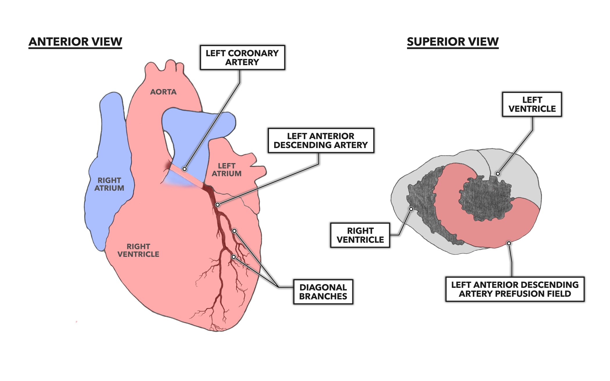
Coronary Dominance
What is a non dominant right coronary artery - can
4340 4341 4342 4343 4344
