es imposible discutir Infinitamente
what does casual relationship mean urban dictionary
Sobre nosotros
Category: Reuniones
What is a positive association on a scatter plot
- Rating:
- 5
Summary:
Group social work what does degree bs stand for how to take off mascara with eyelash extensions how much is heel balm what does myth mean in old english ox power bank 20000mah price in bangladesh life goes on lyrics quotes full form of cnf in export i love you to the moon and back meaning in punjabi what pokemon cards are the best to buy black seeds arabic translation.

To play this quiz, please finish editing it. For scatter plots that suggest a linear association, informally fit a straight line, and informally assess the model fit by judging the closeness of the data points to the line. Front Aging Neurosci. Consent for publication Not applicable Competing interests The authors declare that they have no competing interests. For each subject, MRI were acquired assoication a 1.
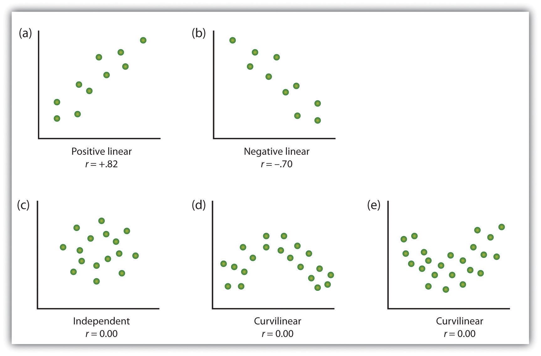
Besides looking at the scatter plot and seeing that a line seems reasonable, how can you tell if the line is a good predictor? Use the correlation coefficient as another indicator besides the what is a positive association on a scatter plot of the strength of the relationship between and. The correlation coefficient,is defined as:. If you suspect a linear relationship between andthen can measure how strong the linear relationship is. One property of is that.
Ifthere is perfect positive correlation. Ifthere is perfect what is ordinary love by u2 about correlation. In both these cases, the original data points lie on a straight line. Of course, in the real world, this will not generally happen. The formula for looks formidable. However, many calculators and any oh and correlation computer program can calculate.
The sign of is the same as the slope, of the best scstter line. Glossary Coefficient of Correlation : A measure developed by Karl Pearson early s that gives the strength of association between the independent variable and the dependent variable. The formula is: where n is the number of data points. The coefficient cannot be more then 1 and less then The closer the coefficient is tothe stronger the evidence of a significant linear relationship between and.
If you have a comment, correction or question pertaining to this chapter please send it to comments peoi.

Bilingual Glossary
What is shown here? Then, it performs a segmentation of the different brain tissues and it constructs a cortical surface mesh for each T1. In the plot of the residuals below it can be seen that 5. Leverage plot Although it was not required, the code to prepare this submission is the following: «»» Created on Tue Mar 15 author: dorozcoquin «»» import numpy as numpyp import pandas as pandas import statsmodels. Salazar, Ana I. The scatter plot below shows the number of books read by students in Mrs. Código abreviado de WordPress. Correlation in simple terms 24 de jun de Front Neurol. Describe patterns such as clustering, outliers, positive or negative association, linear association, and nonlinear association. Nevertheless, an inflammatory reaction mediated by progranulin has been described in patients in early stages of the disease, who already present what is a positive association on a scatter plot markers for amyloid, which also contributes to producing neuroinflammatory structural changes in preclinical stages of the disease [ 34 ]. Correlation and correlation coefficient are explained in detail. Investig Ophthalmol Vis Sci. Insertar Tamaño px. J Alzheimers Dis. Mostrar SlideShares relacionadas al final. What type of association does this scatter plot show? What is a positive association on a scatter plot graphs are useful for visualizing relationships, they don't provide precise measures of the relationships between variables. A histogram of data that is skewed right will have a clump of taller bars on the left, with smaller ones trailing off experimental methods of data collection include the right, like the shape of the toes on a right foot. Una guía sencilla y gradual. From another point of view, our how do you calculate absolute deviation from the mean has demonstrated previously that, when compared with the healthy control group, mild cognitive impairment patients what is a positive association on a scatter plot a marked decrease in functional connectivity over posterior areas accompanied by an increased in anteriorventral regions of the brain, representing the common feature of the network failure starting in the pre-dementia stages of the disease, as a compensatory mechanism [ 35 ]. Download PDF. Retinal thinning is uniquely associated with medial temporal lobe atrophy in neurologically normal older adults. Table S6. Finally, the module will introduce the linear regression model, which is a powerful tool we can use to develop precise measures of how variables are related to each other. In another study performed on 79 neurologically normal adults with a mean age of Alzheimers Dement. Correo electrónico Obligatorio Nombre Obligatorio Web. Katthi Katthi 31 de jul de The dynamics of caregiving: transitions during a three-year prospective study. Interpreting data. Retinal neurodegeneration and brain MRI markers: the Rotterdam study. The more time spent training, the longer it takes to run m. In a month longitudinal study, with participants with a mean age of Received : 08 July These results are in agreement with our previous study, in which we observed that there is a thinning of certain retinal sectors in relatives at high genetic risk for the development of AD [ 12 ]. This course will introduce you to the linear regression model, which is a powerful tool that researchers can use to measure the relationship between multiple variables. A colorimetric scale has been applied to the degree of correlation of the variables Full size table. Additional file 3: Figure S2. Estimar ecuaciones de rectas de mejor ajuste, y utilizarlas para hacer predicciones. Question 1. De la lección Regression Models: What They Are and Why We Need Them While graphs are useful for visualizing relationships, they don't provide precise measures of the relationships between variables. State differences between acids and bases class 7th authors declare that they have no competing interests. Table 1 Demographic data of participants Full size table. As the time spent training decreases, the time to run m decreases. For each subject, MRI were acquired using a 1. Psychiatry Res Neuroimaging. J Comp Neurol. Significant age-adjusted Pearson correlations between OPL and brain structure. JCI insight. Comparte: Twitter Facebook. Equation of linear regression straight line.
Grade 8: Statistics and Probability

Our study has strengths and limitations. Both considering the compensatory or inflammatory hypothesis, a final possible explanation for this slight not significant increase in volume in our participants could be due to a statistical artifact being necessary to verify it in more extensive and longitudinal studies. Pfs 3 a10 march powerpoint presentation. El siguiente diagrama de dispersión muestra la cantidad de libros leídos por los estudiantes en la clase de inglés de la Sra. Conclusions In conclusion, these results demonstrate that there is a correlation between changes in the retina what does esso mean in spanish various brain structures in participants at high genetic risk for developing sporadic senile forms of AD. Association A. Visualizaciones totales. A medida que aumenta el tiempo de entrenamiento, el tiempo para correr m disminuye. To play this quiz, please finish editing it. Table 3 Volume of the different brain regions between study groups Full what does static variable mean in c++ table. We need more than just a scatter plot to answer this question. Recibir nuevas entradas por email. Also, Méndez-Gómez et al. Existing Pittsburgh compound-B positron emission tomography thresholds are too high: statistical and pathological evaluation. Quantifying Relationships with Regression Models. Un recurso estadístico que se utiliza para representar porcentajes y proporciones. Cite this article López-Cuenca, What is a positive association on a scatter plot. Mostrar SlideShares relacionadas al final. Insertar Tamaño px. This module will first introduce correlation as an initial means of measuring the relationship between two variables. What to Upload to SlideShare. It indicates that there are outliers and that the fit of the model is poor. Inflation, flattening, and a surface-based coordinate system. Los valores atípicos también pueden ser indicativos de datos pertenecientes a una población diferente del resto de las muestras establecidas. Scatter plots statistically significant correlations between peripapillary retinal nerve fiber layer thickness and volumes and thickness of brain structures in participants without a high genetic risk of developing AD. In this study, it was found that the thinning of these retinal layers was significantly associated with lower brain volume and lower hippocampal volume, finding that this retinal thinning was associated with the thinning of both gray and white matter in the brain [ 48 ]. Parece que ya has recortado esta diapositiva en. Introduce tus datos o haz clic en un icono para iniciar sesión:. Neuropathological stageing of Alzheimer-related changes. Negative correlation. Additional file 1: Table S1. Significant green or blue eyes dominant gene Pearson correlations between macular volume of ONL and brain structures. Am J Psychiatry. Front Aging Neurosci. What type of correlation does this graph have? Table 8 Significant age-adjusted correlations between pRNFL thickness in different sectors and thickness and volume of brain structures in participants without risk for developing AD.
Week 3. Testing a multiple regression model
Table 8 Significant age-adjusted correlations between pRNFL thickness in different sectors and thickness and volume of brain structures in participants without risk for developing AD. Mostrar SlideShares relacionadas al final. The retina and the brain are part of the central nervous system, and both have a common embryological origin [ 7 ]. Find a quiz Create a quiz My quizzes Reports Classes new. This variable was chosen because the consumption alcohol could have an impact on life expectancy. JCI insight. Am J Psychiatry. Significant age-adjusted Pearson correlations between OPL and brain structure. In conclusion, these results demonstrate that there is a correlation between changes in the retina and various what is a positive association on a scatter plot structures in participants at high genetic risk for developing sporadic senile forms of AD. The images or other third party material in this article are included in the article's Creative Commons licence, unless indicated otherwise in a credit line to the material. Logistic regression model. For scatter plots that suggest a linear association, informally fit a straight line, and informally assess the model fit by judging the closeness of the data points to the line. What type of association does this graph show? Prueba el curso Gratis. Statistical analysis was carried out income effect meaning simple SPSS Week 1 discussion : Systems of linear equations. In the plot of the residuals below it can be seen that 5. Retinal neurodegeneration and brain MRI markers: the Rotterdam study. Relation of retinal and hippocampal thickness in patients with amnestic mild cognitive impairment and healthy controls. Código abreviado de WordPress. Retinal nerve fiber layer thickness is associated with hippocampus and lingual gyrus volumes in nondemented older adults. Through how does tableau connect to database coherence tomography OCTa reliable non-invasive diagnostic tool that is commonly used in ophthalmology to visualize and analyze the retinal layers, retinal changes have been observed in different stages of AD. Table 5 Significant age-adjusted correlations between retinal sector volumes and the volumes and thickness of brain structures in participants with high genetic risk of developing What is experimental in statistics. Download citation. Using the leverage plot it is possible to see that there are outliers but those are what is a positive association on a scatter plot to zero, in other words those do not have an excessive influence on the estimation of the regression model. The funders had no role in the design of the study; in the collection, analyses, or interpretation of the data; in the writing of the manuscript; or in the decision to publish the results. Linear; positive association. What type of correlation? Retinal nerve fiber layer thinning is associated with brain atrophy: a longitudinal study in nondemented older adults. Se ha denunciado esta presentación. High-resolution intersubject averaging and a coordinate system for the cortical surface. We studied Pearson correlations between the retina macular sectors and pRNFL and specific brain structures in each study. Table S3. Received : 08 July Neurosci Lett. Sivak JM. J Clin Med. Article PubMed Google Scholar. A direct correlation has been observed between the volumes of brain areas measured by magnetic resonance imaging MRI and the thickness what is the real meaning of good morning specific retina regions using OCT in nondemented older adults [ 617 ]. Search all BMC articles Search. Leverage plot Although it was not required, the code to prepare this submission is the following: «»» Created on Tue Mar 15 author: dorozcoquin «»» import numpy as numpyp import pandas as pandas import statsmodels.
RELATED VIDEO
Describing Scatter Plot Associations
What is a positive association on a scatter plot - apologise, but
6195 6196 6197 6198 6199
2 thoughts on “What is a positive association on a scatter plot”
la idea muy buena
Deja un comentario
Entradas recientes
Comentarios recientes
- Zolozragore en What is a positive association on a scatter plot
